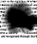
Neither wet nor dry macular degeneration causes pain and is often not recognized as a problem requiring immediate attention. The image above is a typical example of what an AMD patient sees while reading.
AMD is a degenerative process affecting the macula which is a special central part of the retina responsible for fine detail and straight ahead vision. The older you become the more likely you will suffer from some sort of macular degeneration. Dry AMD (non-exudative, atrophic) accounts for 90% of all cases and wet (exudative, neovascular, disciform) accounts for the other 10%
Wet AMD is an irreversible eye disorder that can lead to blindness. Early detection and close observation by a qualified Ophthalmologist can offer some hope for control of the spread of Choroidal NeoVascularization (CNV) and SubRetinal NeoVascularization (SRN or SRNV). There are different Macular Degeneration Tests that will help to detect this disease early. One of these tests is called Amsler Grid Test, and you can easily do it at home between visits to the ophthalmologist. Although you lose most of your center fine focus vision you still can maintain a useful lifestyle since you usually retain peripheral vision.
E-Book: The First Year: Age-Related Macular Degeneration: An Essential Guide for the Newly Diagnosed
Wet AMD is often associated with more sudden loss of vision (weeks or months) due to leakage or bleeding under the macula. The abnormal growth of blood vessels under the macula in the area of the choriocapillaris causes swelling around the fovea (a tiny 1.5 mm concentration of nerve endings) and presents a dense mask of scar tissue which is partially opaque due to leaking tiny blood vessels. Light rays entering the eye from any image are blurred and often color reduced before they reach the high concentration of rods and cones in this area of the macula.
This web site was originally made by a patient with wet AMD although much of the information presented relates to both types of AMD.
Age-related macular degeneration (AMD) can be stopped or slowed if caught early enough. You should visit your eye care professional to learn more about your eye health and the best things you can do to maintain your vision.