The human body is a fantastic thing. There are dozens of organs and systems working in concert to keep you alive and functioning well. Our ancestors developed many traits that helped them survive, and our eyes reflect this ancestry. Our eyes take in information from our environment and then send it to our brains to help us understand what is happening around us. One of the most intricate and beautiful organs is the human eye.
Anatomy of the Eye
The eye is unique in being one of the few organs that we can routinely see. Our eyes work much as a camera does. Let's discuss how the anatomy of the eye impacts how we see.
Everybody's favorite part of the eye is the iris. The iris is the colorful structure of the eye that surrounds the pupil. The iris can expand and contract to regulate how much light is allowed in. As light enters the pupil–the opening in the center of the eye–the iris obscures or reveals the pupil depending on perceived lighting conditions.
The light then passes through the cornea. The cornea is the eye's outermost layer, and it acts as a clear window that lets light into the eye. Behind the cornea is a layer of watery fluid known as the aqueous humor. This fluid has a small amount of protein in it and is secreted by the ciliary body, a muscular structure that helps to support and focus the lens.
Once light passes through the pupil, it strikes the lens. The lens is a transparent structure that focuses light on the retina. Muscles in the eyes called the inferior rectus, and inferior oblique muscles focus the light through the lens. The lens is suspended in position by the suspensory ligaments, also known as Lockwood's ligament.
Behind the lens is a transparent, gelatinous substance known as the vitreous humor. The vitreous humor is mainly made of water and contains some salts, sugars, and collagen fibers. The vitreous humor attaches to various points inside the eye and fills out the central chamber, giving the eye its rounded shape.
Once light has passed through the lens and the vitreous humor, it strikes the retina. The retina is a light-sensitive layer of nerve cells that coats the back of the eye. The retina has an egg-shaped, pigmented area near its center known as the macula. The macula produces high-resolution color images to be sent to the brain and is responsible for our good daylight eyesight.
The macula contains a small pit of closely packed cones known as the fovea. Most people have three different types of cones that break up our color vision into red, green, and blue waves. The fovea is responsible for the detailed focus that the human eye can achieve when performing tasks like reading. Interestingly, the fovea transmits so much data to the brain that about half of the optic nerve is used to transmit information specifically from the fovea.
The whites of our eyes that surround the iris and pupil are known as the sclera. Sclera are an opaque protective layer, mostly made of collagen and elastic fibers.
5 Common Eye Disorders
Some eye disorders are likely to happen more often than others. Like any body part, it is prone to occasional malfunction based on usage or age. "A healthy optic nerve is essential to good vision, yet once damage occurs, it cannot be restored," says Michele Lim, professor of ophthalmology and a glaucoma specialist at UC Davis.
The two most common eye disorders are Myopia (nearsightedness) and hyperopia (farsightedness.) These are straightforward eye disorders that can easily be corrected with glasses or corrective lenses.
But there are more complex problems with the eye. Here are five common eye disorders.
Age-related Macular Degeneration
Age-related macular degeneration (AMD) is the medical term describing the gradual deterioration of vision as people age. As the name implies, this deterioration centers around the macula, the small cluster of cells in the retina that helps us maintain the sharp and high-definition vision we use for tasks like reading and driving.
"Currently, the only treatments for early AMD are over-the-counter antioxidant vitamins that can only slow down but not stop disease progression," says Glenn Yiu, who is a retina specialist and physician-scientist at UC Davis.
Cataract
A cataract is an opacity, or a cloudy layer, that develops over the eye's lens. Cataracts can cause obstructions in a person's vision or make it appear cloudy. In general, cataracts occur in people who are 55 years and older, but they can occur in infants and children in some circumstances.
The lens of the eye has three layers: the capsule, the cortex, and the nucleus. Cataracts can occur in any part of the lens.
There are certain risk factors for cataracts, including smoking, drinking alcohol, family history of cataracts, diabetes, long-term use of some prescription drugs, excessive UV radiation exposure, which can increase the likelihood of developing cataracts.
Glaucoma
Glaucoma is a form of damage to the optic nerve caused by excess fluid. When fluid builds up in the vitreous humour, it can cause increased pressure in the eye, which pushes on the optic nerve causing damage.
There are two forms of glaucoma: primary open-angle and closed-angle glaucoma.
Primary open-angle glaucoma is the most common glaucoma. Normally, the eye drains old fluids out at a slow and steady rate to maintain balance in the eye. In primary open-angle glaucoma, the eye does not drain as it should. Open-angle glaucoma is generally painless at first and is relatively slow to develop.
Closed-angle glaucoma occurs when one's iris is too close to the "drainage angle" in the eye. The iris ends up blocking part of the drainage from the eye, much like an obstruction over a sink drain would.
Diabetic Retinopathy
Diabetes can harm many organs in the body, including the eyes. In diabetic retinopathy, high blood sugar levels cause glucose to accumulate in the tiny blood vessels inside your retina. The high blood sugar causes problems with the blood flow to sensitive parts of your retina. Insufficient blood supply leads to tissue dysfunction and eventually death. Diagnosis of diabetic retinopathy is based on an eye exam.
Corneal Ulcer
Corneal ulcers are sores on the cornea, the outer layer of the eye. These ulcers often come from eye infections but can be caused by problems like dry eye. Corneal ulcers are very dangerous and can cause permanent blindness or permanent eye damage if not treated quickly and effectively.
Symptoms Of An Eye Problem
The best way to protect your vision is to catch eye disorders early when they are most easily treated. It's important to be aware of any changes to their vision. If your vision is becoming more cloudy, blurry, distorted, dulled, or otherwise different from your usual, you should seek the care of an eye doctor.
We've said it before, but we'll say it again: it is important that you regularly see an eye doctor for eye exams. Your regular doctor can perform basic eye tests and examinations, but the eye doctor has specialized training and precise tools.
"As a result, vision loss due to glaucoma can't be recovered. That's why it's important to have regular eye exams that include measurements of eye pressure and assessment of the optic nerve. If glaucoma is recognized early, vision loss can be slowed or prevented." says Lim.
Our eyes are amazing. We depend on them for almost every aspect of our modern lives. Our eyes also give us unique visual experiences from reading books, seeing expressive art and the wonder of visual illusions. Your eyes are an essential but delicate part of your body. You should visit your eye care professional to learn more about your own personal eye health and the best things you can do to maintain it.
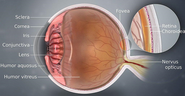
 The optic nerve and the central retinal blood vessels enter the back of the eye at the disc (also called the blind spot). The back 2/3 of the eye is called the retina and gives us our wide field of view vision.
It contains millions of rod and cone cells which convert light energy into electrical signals sent to the brain via the optic nerve. The macula (6x7 mm) is the tiny spot on the retina where finer detail focus occurs. The fovea (1.5 mm) is just behind the macula where the highest concentration of cone photoreceptors are concentrated. Light rays are focused by the lens onto the fovea for straight ahead vision and fine detail. The sclera is the tough outer wall of the eye and the choroid is the thin spongy layer between it and the retina filled with blood vessels.
The optic nerve and the central retinal blood vessels enter the back of the eye at the disc (also called the blind spot). The back 2/3 of the eye is called the retina and gives us our wide field of view vision.
It contains millions of rod and cone cells which convert light energy into electrical signals sent to the brain via the optic nerve. The macula (6x7 mm) is the tiny spot on the retina where finer detail focus occurs. The fovea (1.5 mm) is just behind the macula where the highest concentration of cone photoreceptors are concentrated. Light rays are focused by the lens onto the fovea for straight ahead vision and fine detail. The sclera is the tough outer wall of the eye and the choroid is the thin spongy layer between it and the retina filled with blood vessels.